NYC/NJ Area 1-732-727-0607 (Corporate Office)
Upstate New York Area 1-585-352-6652

VuMAXHD
Simply the BEST. Period.

UNPARALLELED IMAGE QUALITY.
Hands down the gold standard in ophthalmic ultrasound. Unparalled UBM and B-scan image quality with Enhanced Focus Rendering™ and large ultra high resolution screen allows you to capture both crisp still images and record video that can be carefully reviewed frame-by-frame.
ELEGANT.
INTUITIVE.
EXCEPTIONAL.
Elegant user interface provides useful tools that are intuitive, simple, and efficient to use. Time-saving features such as selectable patient database display to easily search and access archive exam records. Document scan orientation with the single click of a button. Replay videos in real-time, slow motion, or frame-by-frame. Super-impose A-scan trace, perform linear and angle measurements, and annotate onto B-scan and UBM images. Auto calculation of axial length average and standard deviation, nine IOL formulas, and lens database for biometric A-scan.


PRESETS
Preset scan modes with settings optimized for
areas of interest
PROBE ORIENTATION
Easy graphical selection
of scan orientation for
all modes
TOOL SET
Simple to use measurement
and annotation tools
REVIEW
Frame-by-frame, full
speed, and slow motion review of video clips
PROBE CHOICES
Choice of 35 or 50 MHz UBM transducers, 12 or 20 MHz B-probes, and immersion or soft-touch A-probes
CONNECTIVITY
Dual-band WIFI, Ethernet, USB, and Bluetooth interface
FLEXIBILITY
Configurable as a B-scan, UBM, A-scan, or in a variety of combinations to meet the needs of your practice
BIG IMAGE
See fine details with high-resolution scans displayed on the 21.5" HD flat panel monitor
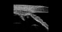
UNPARALLELED UBM.
The VuMAX series from Sonomed Escalon has long been the gold standard in UBM imaging. Now with Enhanced Focus Rendering™ and other enhancements, the VuMAX™ HD delivers the most outstanding UBM images and video clips of the entire anterior chamber.
GLAUCOMA MANAGEMENT
The specialized angle detail scan setting optimizes resolution of different structures at the angle and behind the iris, providing the premier diagnostic tool for identifying causes of glaucoma-related concerns, including angle detail and permeability of the trabecular meshwork, plateau iris syndrome, effects of pupil movement on the angle, and other causes of glaucoma.
ICL SIZING
Accurately measure sulcus-to-sulcus for properly and confidently size ICLs to ensure no post-operative surprises. The specialized sulcus-to-sulcus scan setting is optimally engineered to enable consistent viewing of key anatomical landmarks required to ensure accurate sulcus-to-sulcus measurements.

ANTERIOR SEGMENT
View the entire anterior segment in great detail, with optimized scan settings of the VuMAX™ HD. Clearly visualize the ciliary body, pars plana, and other structures and identify tumors, cysts, trauma, uveitis, and other pathologies. The VuMAX™ HD even allows for visualization and video capture of accommodation.
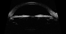
UNPARALLELED B-SCAN.
The all-new B-scan mode of the VuMAX™ HD produces truly outstanding imaging of the posterior segment that has become the new gold standard.

SEE EXTRAORDINARY DETAIL WITH ENHANCED FOCUS RENDERING
Easily visualize extraordinary fine details within video clips and still images generated using proprietary Enhanced Focus Rendering™, providing image quality unmatched by other imaging systems.

OPTIMIZED ULTRASOUND IMAGING SETTINGS
MADE EASY
Select from 4 preset scan modes to optimize image quality in area of interest, including orbit, vitreous body, retina surface, and deep retina / choroid.
QUANTITATIVE ANGLE ANALYSIS.
Easily visualize extraordinary fine details within video clips and still images generated using proprietary Enhanced Focus Rendering™, providing image quality unmatched by other imaging systems.

SULCUS-TO-SULCUS PROBE ALIGNMENT.
Measuring sulucs-to-sulcus is imperative for proper ICL sizing. The key to accurately measuring sulcus-to-sulcus is ensuring proper probe alignment. The VuMAX HD eye tracking feature provides real-time feedback to ensure your scans are properly aligned.
AS YOU LIKE IT.
B-scan. UBM. A-Scan. Any combination.
B-SCAN
Immersion or direct contact A-scan
Manual or automatic capture (cataract, dense cataract, aphakic, silicone oil, and pseudophakic modes)
Auto calculation of axial length, anterior chamber depth, lens thickness, and vitreous, with axial length average and standard deviation provided for up to 10 scans per exam
Standard IOL formulas: Binkhorst, Regression-II, Theoretic/T, Holladay, Hoffer-Q, and Haigis
Post-refractive IOL formulas include Latkany Myopic Regression, Latkany Hyperopic, and Aramberri Double-K
UBM
Unparalleled image quality with proprietary Enhanced Focus Rendering™
12 and 20 MHz frequency transducers available for posterior B-scan
256 ray scan with 2048 sample points for each ray (over a half-million sample points per transducer sweep)
Capture 50-frame video clips at up to 20 frames per second
Store unlimited images and up to 12 video clips in each exam
A-SCAN
Unparalleled image quality with proprietary Enhanced Focus Rendering™
35 and 50 MHz frequency transducers available for UBM
256 ray scan with 2048 sample points for each ray (over a half-million sample points per transducer sweep)
Capture 50-frame video clips at up to 20 frames per second
Store unlimited images and up to 12 video clips in each exam
UBM scan cap kit compatible with ClearScan®device for direct contact UBM scanning